The Complete Dummy's Genetics Guide for Idiots, Part One
the basics
Why do some traits appear in grandparents and grandchildren, skipping the intervening generation? Are there really genes for intelligence, running, mental arithmetic? More broadly, are we in any sense controlled by our genes? How far has our understanding of genetics advanced in recent years, and where is the field heading? What about ethical issues raised by new genetic technologies?
A conceptual understanding of genetics is mandatory for anyone wishing to truly understand questions like these. And, if we don't want to be misled by grandiose or unfounded claims that are regularly made about genetics, we need a solid grasp of how genetics works. This is today more vital than ever, given the ever-accelerating pace of research.
This is the first of a three-part series that collectively aims to cover all the major concepts required for a solid understanding of modern genetics. This article will give you a tour of the basics, with no prior knowledge required. In part two I'll expand on this foundation by covering more complex concepts, and part three will conclude the series by discussing some human-specific areas of genetics in greater detail.
After reading this series, you should have a much more sophisticated understanding of genetics, and you'll be able to utilize this as I plan to write more specialized articles on diseases and other genetics-related stories in the news, several of which will be critiques of a few of the many dubious claims propagated by the media.
Let's start the discussion with three somewhat familiar concepts: cells, genes and DNA.
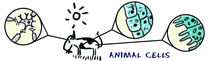
Living things are all made up of cells, self-contained biological units consisting of various components that are themselves made up of nucleic acids, proteins, carbohydrates and fats, all enclosed in a porous membrane. Whether part of an organism consisting of trillions of cells, like a human, or the entire organism as with bacteria and yeast, the cell is the fundamental building block of life.
 An animal consists of many different cell types. Cells of a specific type often cluster together to form tissues. An organ (e.g., the skin or heart) consists of several tissue types. Finally, organ systems are composed of several organs working together (e.g., the nervous system consists of brain, spinal cord, the sense organs and nerves).
Although each cell type is optimized for its own specialized purposes, all animal cells consist of a selection of the same subunits, called organelles, each of which has a specific function (energy production, waste disposal, repair and growth, etc). The cell membrane allows adhesion to neighbouring cells and also communication with the outside world. Cells can receive instructions from outside via specific signalling molecules, electrical impulses, or changes in the conditions within the cell (for example acidity or the concentration of various molecules).
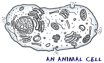 An animal cell consists of an outer membrane and many organelles, suspended in a liquid called cytoplasm. The DNA is stored in the nucleus (the large organelle close to the left side of this cell).
In order to correctly carry out most of the things that it needs to do, a cell requires accurate instructions. These instructions are contained within the genes. Humans each carry around 20,000 genes in almost all of their 35 trillion body cells. The genes collectively contain the information needed to build our bodies and keep them alive.
DNA is the molecule that contains these genes, and it is combined with proteins that help to store and protect it. DNA is organized in such a way that it acts as a code for the production of proteins. It's a very elegant molecule; the code is made entirely from just four different small subunits called nucleotides, connected into a very long, very thin strand of DNA. The nucleotides from one strand pair with those from another strand, to form the familiar double helix shape (see figure). Most of these nucleotide pairs don't do much of anything, but strewn amongst the dead viruses and repetitive sections of junk are the genes, and the switches that control them. A typical gene is a few thousand nucleotide pairs in length, consisting of a specific sequence of the four different nucleotide subunits. It is this sequence that determines which protein(s) the gene can make.
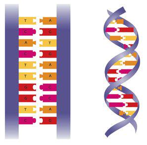 Left: DNA consists of four nucleotides, designated A, C, G and T. Notice that A always bonds with T, and C always bonds with G. Right: the double helix structure of DNA.
Because our bodies consist of many different types of tissues and organs (each with different functions), each body cell that makes up these tissues and organs needs different proteins at different times. So we can't just have all our genes busily making proteins around the clock; our genes need to be regulated – they need a way to produce certain proteins in certain amounts and at certain times. And there's a lot to keep track of: our genome – the entirety of our DNA – has over 3 billion nucleotide pairs. You could fit about 30 million pairs into 1 centimetre (75 million pairs per inch). If you stretched out the DNA in one of your cells, it would be around 2 metres (6 feet) long. Multiply this by the number of cells in your whole body, and you'd have enough DNA to stretch to the Sun and back over 50 times! And if we multiply that by the 7 billion people on the planet, then we'd get enough human DNA to stretch out of our solar system and past all the stars we can see in the night sky. In fact, all this DNA would be able to leave our own galaxy and reach into a neighbouring one.1
How do our bodies take care of and organize all this DNA? Within each body cell a genome is packed into a central structure called the nucleus (see figure). The sheer volume of genetic material, combined with the necessity that most genes be switched off most of the time, means that the usual state of our DNA is to be kept tightly packed, folded, rolled and scrunched up. This is accomplished using proteins, in a DNA-protein complex called chromatin. The chromatin is kept compact and stops the genes from making new proteins when they shouldn't. On those occasions when a gene is called upon to go to work, the section of chromatin containing the gene in question is unfurled, unpacked, unwound and unscrunched long enough for it to make the requisite amount of protein, after which it's bundled back up and packed away like an insane granny in the attic.
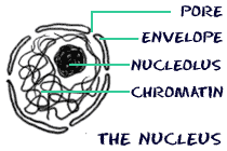 The nucleus is the largest organelle in the cell. The nuclear envelope has pores to allow the transfer of particles into and out of the nucleus. The chromatin consists of DNA wrapped in various proteins. The nucleolus is a genomic region involved in the production of ribosomes, small cellular machines that reside outside the nucleus and convert genetic information into proteins.
As well as being tightly packed, our DNA (in the form of chromatin) is also divided into 23 pairs of chromosomes. We inherit one of each pair from each of our parents, giving us two of each chromosome. For example, a child's mother has two copies of chromosome 1, as does their father. This child got one of their mother's chromosome 1, and one of their father's. The first 22 pairs of chromosomes are known as autosomes, and thus we have two copies of genes on any of our autosomes – one from each of our parents.
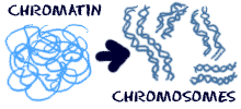 Inactive chromatin is highly condensed and divided into 23 pairs of chromosomes.
The 23rd pair of chromosomes is different; these are the sex chromosomes. The most important function of the sex chromosomes is – you guessed it – determining a person's sex. Females have two X chromosomes, while males have an X and a Y. Everyone inherits one of their mother's two X chromosomes, but girls inherit a second X, from their father, while boys inherit their father's Y chromosome. It is thus the father who determines the sex of his children. A runt among chromosomes (see figure), the Y has very few genes, and those that it does have are almost exclusively devoted to producing maleness in their owners.
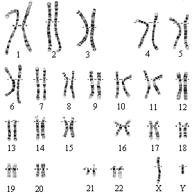 The 23 pairs of human chromosomes, imaged in a karyogram. These are from a male (you can see that the 23rd pair of chromosomes (which are the sex chromosomes) consists of an X and a Y).
So now we know how our genetic material is organized and that the genes make proteins. We also know that a gene consists of a specific sequence of nucleotides that determines the protein it will produce, but how does this work? As we learned already, the chromosomes are safely stored in a central, cordoned-off area of a cell called the nucleus. When a cell needs to make a protein, it duplicates the relevant section of DNA by copying the sequence into a molecule called messenger RNA (mRNA). mRNA is single-stranded and disposable, and it moves out of the nucleus through pores in the nuclear envelope, where its code is read and interpreted by ribosomes that produce the required protein, after which the mRNA is recycled. The DNA is like the original, master copy of a vital document, and so is kept safe inside the nucleus. mRNA is a transcribed copy of a specific section of the master document, and so can be removed, read and thrown away as required.
But how does the cell control which genes are operative at a given time? In addition to the genes, and usually near to them, DNA also contains sequences of nucleotides that act as switches, which can be turned on or kept off by specific regulatory molecules (proteins or regulatory RNA strands). This regulation can occur in one or more of several different ways: a cell can keep the DNA tightly packed and inactive, or open it up for transcription using structural or chemical signals; it can control the timing and duration of mRNA production from opened DNA using regulatory switches and proteins (or even other types of RNA); it can modify transcribed mRNA, perhaps rearranging it to form different sequences and so make different proteins; and it can send signals to increase or decrease the number of copies of a specific mRNA molecule, to determine how much protein an mRNA produces.
All these mechanisms (and more which we won't consider here) work in concert to regulate protein production with great precision. In this way, each cell in our body can produce different proteins at different times, as its circumstances dictate.
There's endless wear and tear involved in being alive, and up until adulthood our bodies are constantly growing. Even as adults, throughout our bodies cells are continuously dying or being killed, and must be replaced. Around 2 million red blood cells die per second; if you've ever donated a pint of blood, you lost around 2.5 billion red blood cells (plus many other types of cells). All our other body cells also wear out and die, and can also succumb to injury. A child starts out as a single fertilized egg cell, and within a couple of decades has become an adult of 35 trillion cells. All this means that large multicellular creatures like us must produce a huge number of new cells each day.
A new cell comes from an existing cell that divides into two. We learned earlier that our cells each carry a copy of our entire genome. So before a cell can divide into two new cells (and in addition to replicating all its other components), it must make an extra copy of the genome so that both daughter cells can each have one. This requires the unwinding and copying of every one of the 3 billion nucleotide pairs. Because DNA is double-stranded, it can be separated into its two constituent strands. Each single strand can then be used as a template to make the complementary strand, producing two identical double-stranded daughter molecules of DNA.
How does this work? Recall from an earlier figure that there are four nucleotides (A, C, G and T), and that an A from one strand always bonds to a T from the complementary strand, and likewise C always bonds with G. This means that when the cell unwinds the DNA double helix, it can look at each single strand of DNA and will always know how to build the other, complementary, strand. For example, if the next nucleotide on a template strand is an A, the cell will add a T to the complementary strand. This elegant process of DNA replication, though it has amazing fidelity, is still prone to the occasional mistake; these mistakes are called mutations, and we'll talk about these and their implications another time.
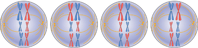
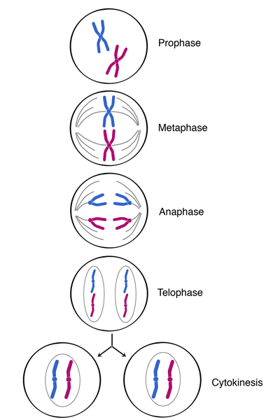 Cell division (simplified only to include one pair of chromosomes). Before cell division can occur, each chromosome must duplicate itself into two sisters, connected to each other near the centre, as has already occurred here (Prophase). The chromosomes are then aligned along the central plane of the cell (Metaphase), using tiny tubes. The sisters then move down the tubes to opposite poles of the cell (Anaphase), after which two new nuclei form around the separated chromosomes (Telophase) and the cell physically divides in two new daughter cells, with one sister from each pair in each (Cytokinesis).
We know that a human starts out as a single cell that got one copy of each chromosome from each parent. If you think about this for a second, you'll realize that what we've discussed so far concerning cell division won't work for sexual reproduction: if sex cells had the same amount of genetic information as other body cells, an offspring would end up with twice the amount of DNA as its parents, and this doubling of DNA would compound in each generation.
So an egg cell and a sperm cell must have half the number of chromosomes of our other body cells (i.e., a single copy of each chromosome, rather than a pair of each). How does this halving work? Normally, each chromosome is copied before cell division occurs, so the chromosome number doubles prior to cell division and is halved during cell division, leaving the original number of chromosomes in the daughter cells.
But during cell division for the production of eggs and sperm (a process known as meiosis), this duplication is followed by two rounds of cell division, leading to an overall halving of the number of chromosomes that end up in the egg or sperm cell.2 Each chromosome is present as a singleton, rather than being one of a pair. So when a sperm cell fuses with an egg, the single chromosomes contributed by each cell combine into pairs, ensuring the embryo has the correct amount of DNA.
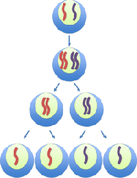 Egg/sperm production. Top: one of the 22 pairs of homologous autosomes in a cell, the red member of the pair inherited from one parent, the blue one from the other. Next: each chromosome of the pair is duplicated. Bottom half: two rounds of cell division first take the chromosome number back to normal, then finally produce four (egg or sperm) cells, each with half the number of chromosomes. When a sperm cell fuses with an egg, the genetic material combines to again produce a cell with a complete set of chromosomes.
Meiosis includes a couple of processes that serve to greatly increase genetic variation in offspring. These both occur during the first of the two rounds of cell division, after the chromosomes have duplicated. The first occurs due to the random inheritance of one parental chromosome from each of the 23 pairs. When the chromosomes line up prior to the cell dividing, the cell does not check which parental chromosome is on which side of the dividing line, leading to a randomization of chromosomes in the two daughter cells. This means that, on average, siblings will share (a random) half of their genetic material with each of their brothers or sisters.
 If we had only 3 pairs of chromosomes, there would be 23 = 2 x 2 x 2 = 8 ways to divide them between daughter cells. Here we see four different possible initial alignments along the central vertical axis, as used for cell division; you can see the eight different possible combinations that could occur (after the left-hand chromosomes have separated from the right-hand ones). Since we actually have 23 pairs of chromosomes, there are in fact 223 = 8.4 million possible arrangements.
The second process that increases genetic variation is a mixing-up of the alleles by swapping sections of chromosomes, known as crossing over (see figure). Because the two chromosomes are analogous to one another along their entire length (they contain different versions of the same genes at any given position), variability can be increased by breaking the chromosomes and exchanging one or more sections between them. The resulting unique chromosomes later go their separate ways, each ending up in a different gamete (egg or sperm cell).
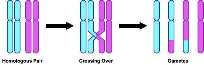 Left: Each of the chromosomes inherited from a parent have been duplicated into a homologous pair (the second step in the above figure on egg/sperm production). Centre: two of the homologues exchange sections. Right: the resultant four chromosomes are divided between four gamete cells, each with differing genetic material.
Together, these two mechanisms can produce almost unlimited shuffling of genes, which is why siblings usually look similar, but can be strikingly different in certain characteristics.
In part two of the series, we'll consider the effect that variations in genes have on individuals, look at how some well-known traits work, and examine some more complex situations such as how the outside world interacts with our genes and the effect this has upon us.
We won't go into the calculations that include all living things on Earth; it's sufficient to state that all that DNA would comfortably stretch around the entire known universe dozens of times.
There is an initial doubling of the genome, followed by two halvings, thus leaving half the original, undoubled, amount of DNA (1 —> 2 —> 1 —> 0.5).